STUDY TITLE: Common Genetic Variation Indicates Separate Causes for Periventricular and Deep White Matter Hyperintensities
SUMMARY: Discovery of 11 genomic regions associated with periventricular white matter hyperintensities that indicate brain lesions.
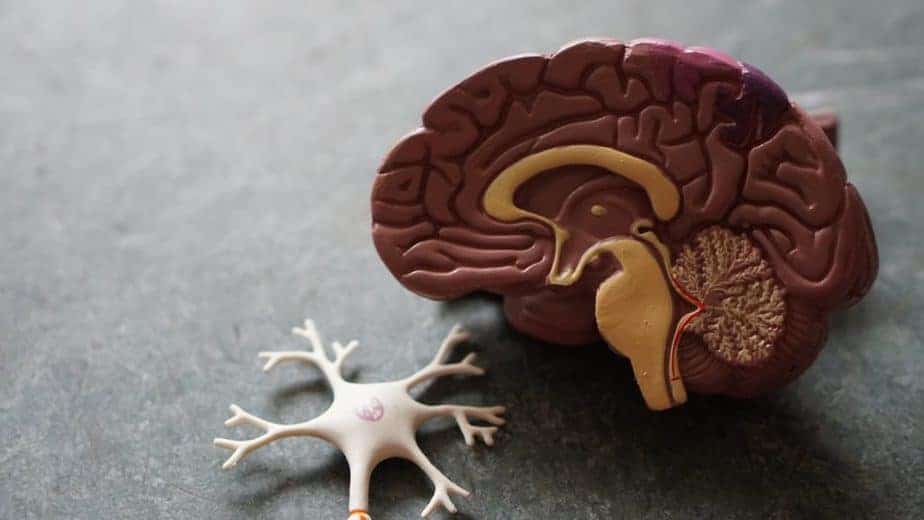
OVERVIEW: The brain is a delicate organ that requires constant blood flow. Strokes occur when a large part of the brain is no longer supplied with blood due to a major clot or a bleed – this leads to obvious brain damage. However smaller clots or bleeds might kill or damage brain cells, possibly causing dementia or movement issues. These might go undetected until the patient gets a brain scan: the damage shows up as a brighter white spot, called a hyperintensity. This study looked for these white spots in brain scans near the fluid sacs in the center of the brain, called ventricles. These brain lesions are called periventricular white matter hyperintensities. By looking at brain scans from over 26,000 people, the authors identified 11 genomic regions associated with periventricular white matter hyperintensities. The presence of these lesions was associated with an increased risk of stroke but was not associated with an increased risk of dementia.
DID YOU KNOW? There is some evidence that white matter lesions may be preventable by not smoking and preventing hypertension, type 2 diabetes, and obesity. [SOURCE]
SAMPLE RESULTS: Learn more about the Nebula Research Library.
BRAIN LESION-ASSOCIATED VARIANTS: rs3744020, rs3758575, rs12928520, rs275350, rs7596872, rs72934583, rs57242328, rs7213273, rs1993484, rs11838776, rs1799983
ADDITIONAL RESOURCES:
What is white matter disease? (Video)
White matter disease
YOU MAY ALSO BE INTERESTED IN:
Stroke (Malik, 2018)
Cerebral cortex thickness (Grasby, 2020)
Total brain volume (Zhao, 2019)
WEEKLY UPDATE: June 25, 2020